
MCL Injuries in the Setting of ACL Reconstruction
Anterior cruciate ligament (ACL) tears rarely happen in isolation. A significant number of patients who tear their ACL also injure the medial collateral ligament (MCL) — usually from the same twisting or valgus (knock-knee) force that damaged the ACL in the first place. Understanding how these two injuries interact is essential for getting the treatment sequence right, because managing a combined ACL-MCL injury is not simply "fix both ligaments at once."
Why the two injuries travel together
The MCL runs along the inner aspect of the knee and is the primary restraint against valgus stress (the knee buckling inward). The ACL, meanwhile, resists anterior tibial translation and rotational forces. A classic mechanism — a planted foot with the knee forced inward, as seen in skiing falls, football tackles, or basketball pivots — loads both structures simultaneously. This is sometimes referred to as part of the "unhappy triad" when the meniscus is also involved.
Grading the MCL injury
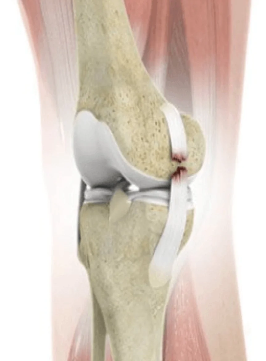
MCL injuries are graded on a I–III scale based on the degree of laxity when the knee is stressed in slight flexion:
Grade I: Mild sprain, minimal laxity (0–5 mm of gapping), ligament fibers intact
Grade II: Partial tear, moderate laxity (5–10 mm), some fiber disruption
Grade III: Complete tear, significant laxity (>10 mm), no firm endpoint on stress testing
Grading matters enormously here, because it drives the treatment algorithm more than the ACL injury itself does.
The core treatment principle: let the MCL heal first
Unlike the ACL, which has poor intrinsic healing potential due to its intra-articular location and limited blood supply, the MCL is extracapsular and has an excellent blood supply. Grade I and II MCL injuries heal well with non-operative management: bracing, protected weight-bearing, and progressive range-of-motion exercises.
This leads to the standard approach for combined injuries:
Brace and rehab the MCL first, typically for 4–6 weeks, allowing it to scar down and regain stability.
Reassess laxity once swelling and pain subside and range of motion is restored.
Proceed with ACL reconstruction once the MCL has healed and the knee has regained motion and reduced inflammation.
When does the MCL need surgery?
Isolated MCL surgical repair or reconstruction is uncommon, but it's considered in specific situations:
High-grade Grade III tears with gross instability that doesn't improve with bracing
Combined injuries with a "Stener-like" displacement or tissue interposition preventing healing
Chronic MCL insufficiency with persistent valgus instability, often in revision ACL cases
Multi-ligament knee injuries (e.g., ACL + MCL + posterolateral corner)
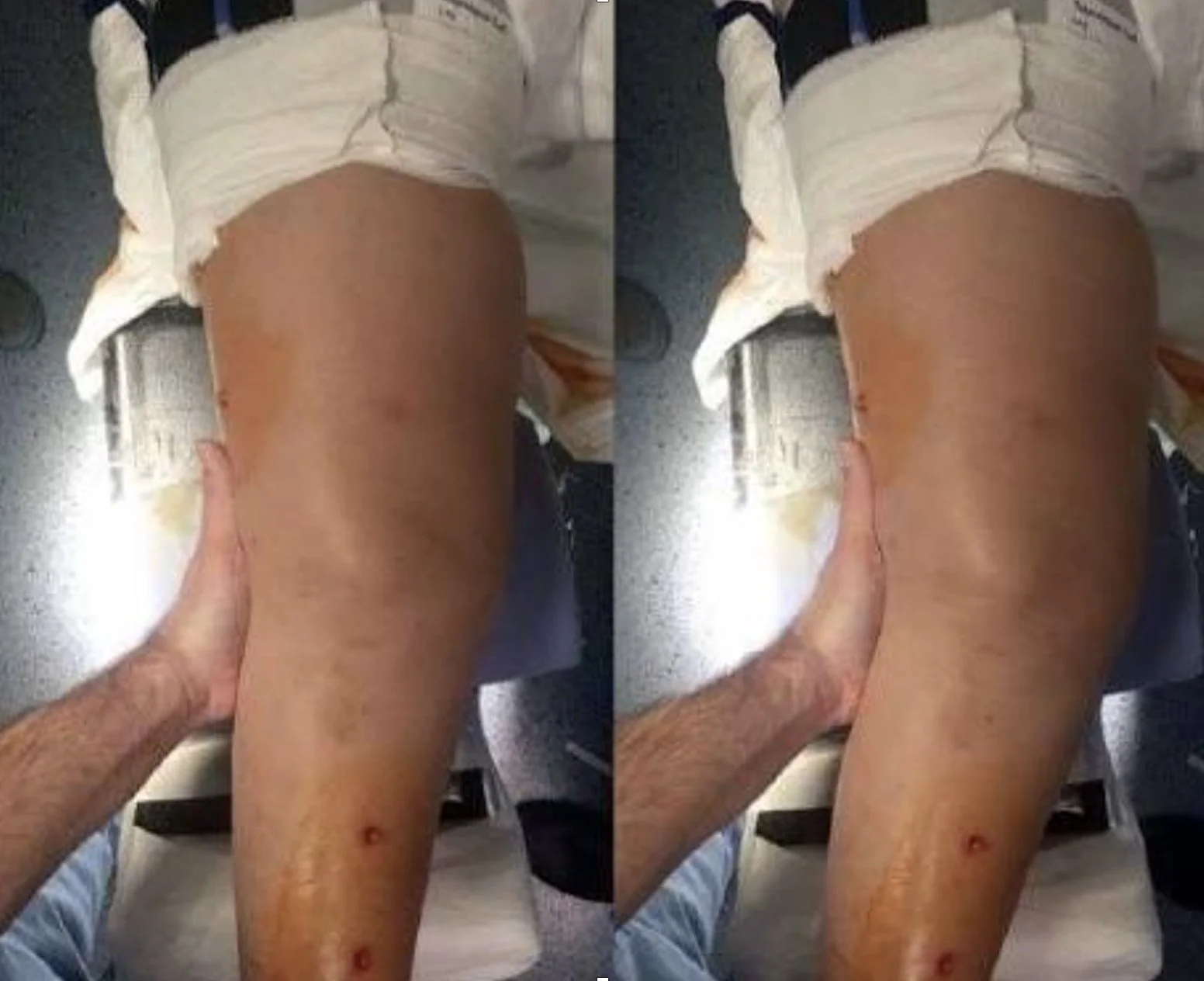
Anteromedial Rotatory Instability (AMRI)
One reason surgeons scrutinise the MCL so closely in ACL-injured knees is a specific instability pattern called anteromedial rotatory instability (AMRI). This occurs when the superficial MCL, deep MCL, and posteromedial capsule are compromised alongside the ACL, allowing the medial tibial plateau to rotate and translate anteriorly relative to the femur.
Clinically, AMRI is picked up through an increased anteromedial drawer test — with the tibia externally rotated, excessive anterior translation of the medial tibial plateau signals combined ACL and medial-sided (MCL/posteromedial corner) insufficiency, rather than an isolated ACL tear.
Why it matters:
Missed AMRI is a cause of ACL graft failure. If significant medial-sided laxity is overlooked and only the ACL is reconstructed, the graft is left absorbing abnormal rotatory and valgus loads it wasn't designed to resist, predisposing it to stretch out or fail.
In short, AMRI is the clinical fingerprint surgeons look for to confirm that the MCL and posteromedial structures — not just the ACL — need to be accounted for in the treatment plan.
Why timing matters for outcomes
Studies have generally supported the staged approach over simultaneous reconstruction of both ligaments in most Grade I–II, largely because:
Combined acute surgery on a swollen knee correlates with higher rates of stiffness
The MCL's superior healing capacity makes surgery for it often unnecessary
That said, an unstable, gapping MCL left unaddressed during ACL reconstruction can compromise the ACL graft over time by exposing it to abnormal valgus loads — so surgeons do pay close attention to residual MCL laxity at the time of ACL surgery, even after a bracing period.
Rehabilitation considerations
Rehab for combined injuries tends to progress more conservatively in the early phase:
Hinged knee bracing to limit valgus stress
Early emphasis on quadriceps activation and range of motion to avoid stiffness
Delayed initiation of aggressive lateral and pivoting drills compared to isolated ACL rehab
Close monitoring for persistent valgus laxity, which may signal incomplete MCL healing
The bottom line
Combined ACL-MCL injuries are common, but the MCL's excellent healing capacity means most cases don't require surgery on that ligament at all. The prevailing approach — bracing and rehabbing the MCL first, then reconstructing the ACL once the knee is calm and stable — tends to produce better range-of-motion outcomes than tackling both ligaments simultaneously. Surgical MCL treatment is reserved for higher-grade instability, multi-ligament injuries, or cases where conservative care fails.
