
Strength Recovery After ACL Reconstruction

01 · Why Strength is the Metric that Matters
After an ACL reconstruction, strength restoration can take 6-9 months, and it is the single most modifiable factor standing between a patient and a safe return to sport.
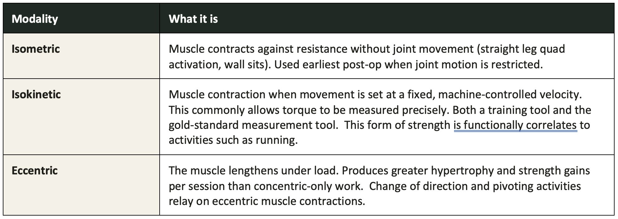
02 · Strength is Not a Single Entity
There are numerous forms of strength that need to be rehabilitated and even quantified after an ACL reconstruction.
03 · Muscle Contraction Control: Why the Nervous System Matters as Much as the Muscle
Strength, in the narrow sense of maximum force output, tells only part of the story. Equally consequential is the nervous system’s capacity to control that force — the timing, gradation, and completeness with which a muscle can be voluntarily activated on demand. Following ACL reconstruction, the quadriceps in particular are subject to arthrogenic muscle inhibition (AMI), a reflexive, presynaptic suppression of motor neuron excitability triggered by joint effusion, pain, and altered mechanoreceptor input rather than by any structural weakness of the muscle itself [13]. AMI can prevent complete voluntary activation of the quadriceps even when the muscle retains its underlying force-generating capacity, and it has been shown to persist well beyond the acute postoperative period in a substantial proportion of patients [13].
This distinction carries real clinical weight. A patient can register an encouraging value on a dynamometer while still being unable to reliably recruit that strength during the split-second demands of cutting, landing, or deceleration, precisely the movements implicated in the original injury. Rehabilitation strategies that address contraction quality directly, through neuromuscular electrical stimulation, biofeedback, and exercises that demand rapid and controlled force production rather than only maximal effort, are therefore targeting a distinct and clinically meaningful dimension of recovery, one that a peak strength number alone does not capture.
04 · Open Chain and Closed Chain: A Historical Caution the Evidence Has Overturned
Exercises are broadly classified by whether the distal segment of the limb is fixed or free. In a closed-kinetic-chain (CKC) exercise, such as a squat, leg press, or step-up, the foot remains planted against a stable surface while force is transmitted through the entire limb; in an open-kinetic-chain (OKC) exercise, such as a seated knee extension, the distal segment moves freely against resistance while the proximal segment stays fixed. For much of the modern history of ACL rehabilitation, OKC quadriceps exercises — particularly knee extension performed near terminal extension — were withheld in the early postoperative period, out of concern that the isolated quadriceps contraction would generate anterior tibial shear force and place undue strain on the healing graft. This caution traces to early biomechanical work demonstrating greater anterior tibial displacement during OKC exercise than during CKC exercise in the ACL-deficient knee, a finding that shaped clinical practice for a generation [14].
Subsequent randomized controlled trials and systematic reviews have substantially revised this picture. Restricted-range OKC exercise introduced early in rehabilitation has not been shown to compromise graft integrity, increase anterior tibial laxity, or impair functional outcomes relative to CKC-only protocols, and a systematic review and meta-analysis directly comparing the two approaches found no clinically meaningful difference in laxity between groups regardless of when OKC exercise was introduced [7, 15]. What the original caution overlooked is that CKC exercise alone does not adequately isolate or load the quadriceps, particularly in a knee guarding against pain and effusion, and can permit compensatory reliance on the hip and posterior chain rather than restoring genuine quadriceps strength [6]. Judiciously dosed OKC exercise, introduced early and progressed through a controlled range of motion, is now recognized as an important tool for directly targeting quadriceps strength and addressing the activation deficits described above, rather than a risk to be managed by avoidance [7].
05 · How the Graft Itself Shapes Recovery
The graft is not a neutral bystander in this process. Whichever tendon is harvested to rebuild the ACL leaves a measurable strength deficit at its donor site and that deficit follows a different timeline for every graft type. Patella and quadriceps tendon will leave increased deficits in the extension mechanism of the knee while hamstring tendons will leave some residual weakness to knee flexion. However, with a good rehabilitation program, most of these defects can be reduce.
The reality is, even after a smooth recovery, most patients will have some quadriceps weakness after full ACL reconstruction rehabilitation.
06 · Beyond the Knee: Hip Abductors and Core

The ACL doesn't fail in isolation from the rest of the kinetic chain. A weak hip or an uncontrolled trunk changes how load arrives at the knee before the quadriceps or hamstrings ever get involved. Hip abductor and external rotator strength help prevent femoral internal rotation and knee valgus during cutting, landing, and deceleration, the same movement patterns implicated in the original ACL injury. Core endurance stabilizes the trunk over the base of support, reducing the compensatory movement strategies that can increase load on the knee.
07 · Measuring Strength Objectively
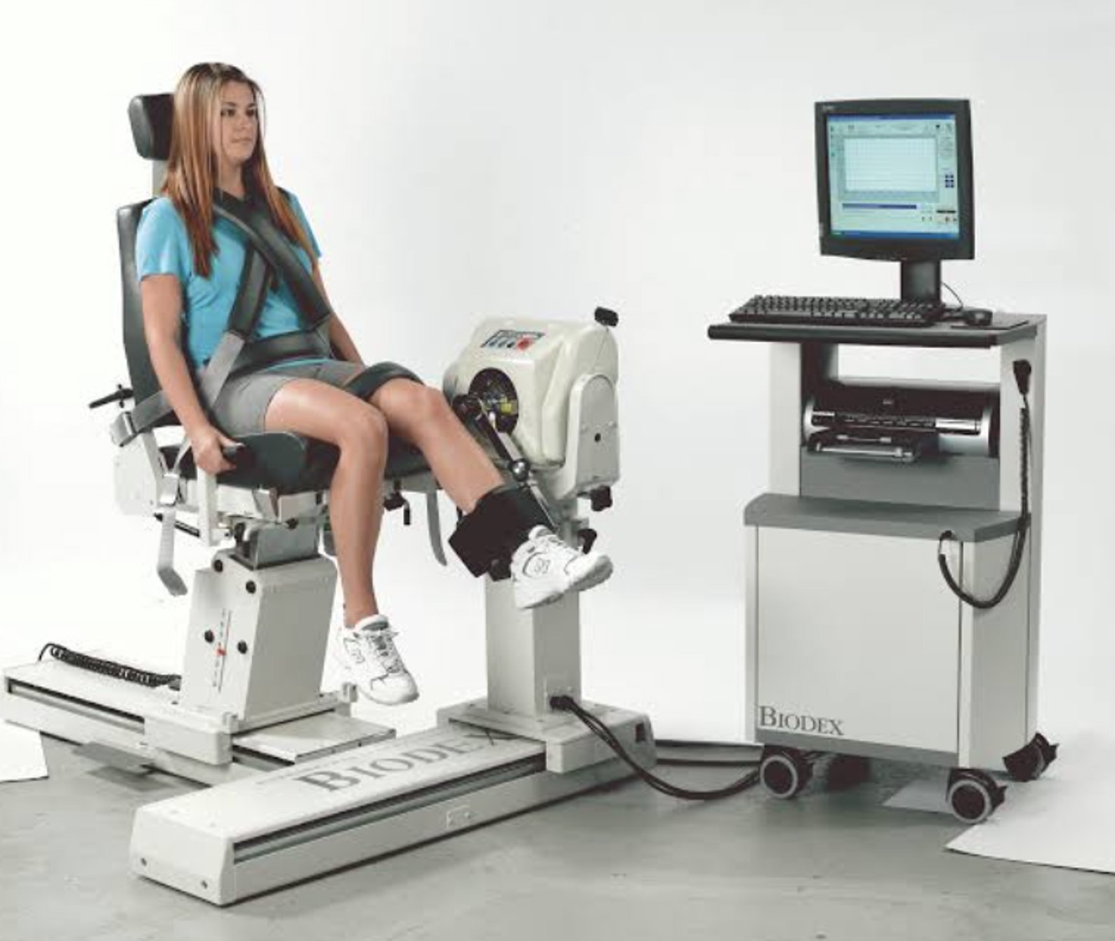
Isokinetic dynamometry, a motorised device that controls movement velocity while measuring torque throughout the range of motion, is treated as the reference standard for quantifying quadriceps and hamstring strength after ACL reconstruction. Strength is most often expressed as a limb symmetry index (LSI): the surgical limb's strength as a percentage of the uninjured limb's strength. A quadriceps strength index of 90% or greater is the threshold most consistently used to define readiness in the return-to-sport cohorts discussed below.
Where a dynamometer isn't available, hand-held dynamometry with fixed stabilization, or a one-repetition-maximum knee extension test, are used as practical alternatives — though isokinetic testing remains the more precise and more extensively validated option.
08 · Does Strength Actually Predict Return to Sport — and Fewer Graft Tears?
Strength is not an isolated metric that ensures fewer graft ruptures. However, some studies have shown that it likely matters when it is used as a metric along with other quality of movement and performance testing batteries, such as single leg hopping. Unfortunately, the exact testing metrics with the highest correlation with safe return to sport has not been established. In addition, most return to sport protocols only evaluated peak forces to isometric and isokinetic strength. We currently do not know how quality of strength during full range of motion or rehab quality of eccentric strengths affects the ability to return to sport and prevention of graft injury.
Sources
[1] Grindem H, Snyder-Mackler L, Moksnes H, Engebretsen L, Risberg MA. Simple Decision Rules Can Reduce Reinjury Risk by 84% After Anterior Cruciate Ligament Reconstruction: The Delaware-Oslo ACL Cohort Study. Br J Sports Med, 2016 [Level II]
[2] Kyritsis P, Bahr R, Landreau P, Miladi R, Witvrouw E. Likelihood of ACL Graft Rupture: Not Meeting Six Clinical Discharge Criteria Before Return to Sport Is Associated With a Four Times Greater Risk of Rupture. Br J Sports Med, 2016 [Level II]
[3] Autograft Type Affects Muscle Strength and Hop Performance After ACL Reconstruction: A Randomised Controlled Trial Comparing Patellar Tendon and Hamstring Tendon Autografts. Knee Surg Sports Traumatol Arthrosc, 2021 [Level I]
[4] Weaker Quadriceps Muscle Strength With a Quadriceps Tendon Graft Compared With a Patellar or Hamstring Tendon Graft at 7 Months After Anterior Cruciate Ligament Reconstruction. American Journal of Sports Medicine [Level III]
[5] Athletes With Bone-Patellar Tendon-Bone Autograft for ACL Reconstruction Were Slower to Meet Rehabilitation Milestones and Return-to-Sport Criteria Than Athletes With Hamstring Tendon Autograft or Soft Tissue Allograft: Secondary Analysis From the ACL-SPORTS Trial. J Orthop Sports Phys Ther, 2020 [Level III]
[6] Mikkelsen C, Werner S, Eriksson E. Closed Kinetic Chain Alone Compared to Combined Open and Closed Kinetic Chain Exercises for Quadriceps Strengthening After Anterior Cruciate Ligament Reconstruction With Respect to Return to Sports: A Prospective Matched Follow-up Study. Knee Surg Sports Traumatol Arthrosc, 2000 [Level II]
[7] Fukuda TY, Fingerhut D, Moreira VC, et al. Open Kinetic Chain Exercises in a Restricted Range of Motion After Anterior Cruciate Ligament Reconstruction: A Randomized Controlled Clinical Trial. American Journal of Sports Medicine, 2013 [Level I]
[8] Isokinetic Eccentric Training Is More Effective Than Constant Load Eccentric Training for Quadriceps Rehabilitation Following Anterior Cruciate Ligament Reconstruction: A Randomized Controlled Trial. Randomized controlled trial [Level I]
[9] Cross-Education Improves Quadriceps Strength Recovery After ACL Reconstruction: A Randomized Controlled Trial. Randomized controlled trial [Level I]
[10] A Randomized Controlled Trial of Eccentric Versus Concentric Cycling for Anterior Cruciate Ligament Reconstruction Rehabilitation. Randomized controlled trial [Level I]
[11] The Effects of Core Stabilization Exercises on the Neuromuscular Function of Athletes With ACL Reconstruction. Scientific Reports [Level I]
[12] Barfod KW, Feller JA, Hartwig T, Devitt BM, Webster KE. Knee Extensor Strength and Hop Test Performance Following Anterior Cruciate Ligament Reconstruction. Knee, 2019 [Level III]
[13] Hart JM, Pietrosimone B, Hertel J, Ingersoll CD. Quadriceps Activation Following Knee Injuries: A Systematic Review. Journal of Athletic Training, 2010 [Systematic Review]
[14] Yack HJ, Collins CE, Whieldon TJ. Comparison of Closed and Open Kinetic Chain Exercise in the Anterior Cruciate Ligament-Deficient Knee. American Journal of Sports Medicine, 1993 [Level II]
[15] Perriman A, Leahy E, Semciw AI. The Effect of Open- Versus Closed-Kinetic-Chain Exercises on Anterior Tibial Laxity, Strength, and Function Following Anterior Cruciate Ligament Reconstruction: A Systematic Review and Meta-analysis. Journal of Orthopaedic & Sports Physical Therapy, 2018 [Level I]
This article summarizes primary orthopaedic and rehabilitation research restricted to Level I–III study designs (randomized controlled trials, prospective comparative cohorts, and retrospective comparative cohorts). It is not medical advice and does not replace an individualized rehabilitation program supervised by a physical therapist or surgeon.
